Retroesophageal right subclavian artery in Thai: A case report
Abstract
Background : The variable structures and pattern of the organs in the body are normally found during dissection of formalin-embalmed cadavers in the practical class of Gross Anatomy. The variation of the right subclavian artery is one of the variable structures. Normally, this artery is a branch from brachiocephalic trunk, which is the first branch of the arch of aorta. The observation on mediastinum dissection of the male cadaver, 89 years of age in the Gross Anatomy Laboratory, Department of Anatomy, Faculty of Medicine, Khon Kaen University, it has been found that the right subclavian artery is a branch arising from posterior aspect of the distal part of the arch of aorta, it runs behind esophagus and directly to the right upper extremity, called retroesophageal right subclavian artery (RRSA). This finding is the first case in our laboratory since the year 1997 to 2004 totalled 480 cadavers. The variable of the RRSA is highly investigated in case of abnormal development of the arch of aorta thus, the surgeon and related physician should be always aware. Objectives- Describe the anatomical characteristics of the RRSA in these aspects: the position of the origin, the diameter, the length and the course of this artery.
2. Describes the surrounded structures related to this
artery.
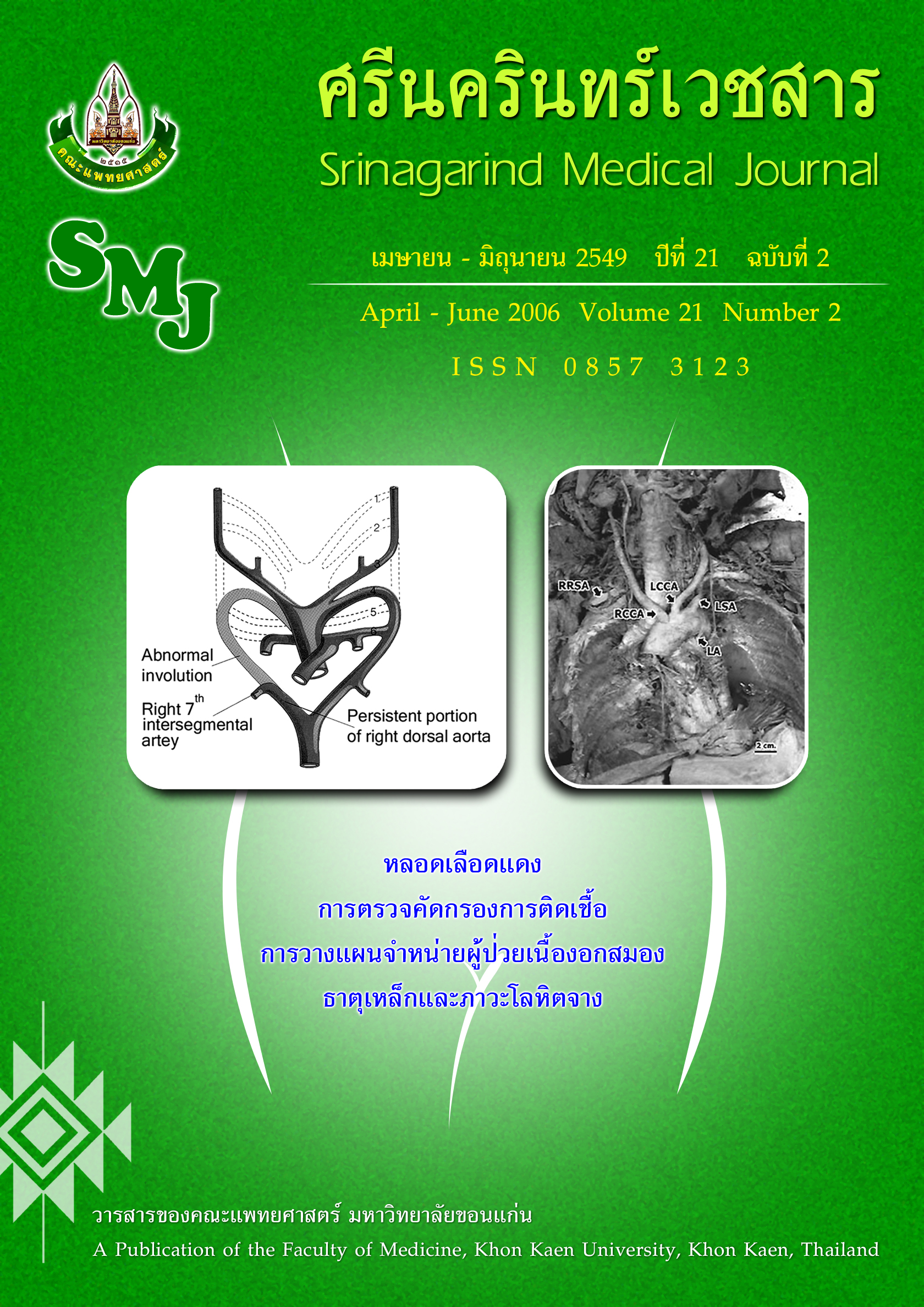
Result : The observation while carefully dissecting the mediastinum of the male cadaver, 89 years old of age. It had been found that the brachiocephalic trunk is absent. The first branch of the arch of aorta is the right common carotid artery, the second is the left common carotid artery, the third is the left subclavian artery and the fourth branch is the RRSA. The RRSA arises from the posterior of the distal part of the arch of aorta, next to the left subclavian artery. The course of this artery is behind the esophagus traversing the mediastinum to supply the right upper extremity. However, there is no variable of the branches from RRSA. Measurement with the digital vernier caliper was done and revealed the length of the RRSA is 8.86 centimeters, the diameter at the proximal and distal part of RRSA are 0.99 and 0.83 centimeters respectively. Furthermore, from this study, the right recurrent laryngeal nerve does not hook around the RRSA as normal.
Conclusion : The RRSA is a defective development of the right fourth aortic arch and/or part of the right dorsal aorta, a segment between the right fourth aortic arch and the right seventh intersegmental artery which may be degenerated during embryological development. This is the first report on the RRSA in Thai. The incidence of this anomaly in Thai should be further researched and accumulated.